


Mangostan - Die Gesundheitsfrucht für Diabetiker
Die 54 jährige Tschechin Ivana Subrt leidet seit 3 Jahren an Entzündungsschüben durch eine Borreliose-Erkrankung sowie an einem gestörten Insulinhaushalt.
„Bisher hatte ich immer schlechte Blutsenkungswerte, leider auch schlechte Blutzuckerwerte und darüber hinaus auch Hautprobleme. Seit 6 Wochen nehme ich täglich 2x 30ml antioxidativen Mangostan-Saft mit zusätzlich angereicherten Naturstoffen für den Zellschutz zu mir. In dieser Zeit habe ich alles andere weggelassen, meine Antibiotika und auch meine Zuckertabletten. Unglaublich, aber wahr: Mein Blutsenkungswert hat sich von 75 mm auf 15 mm verbessert. Der Blutsenkungswert zeigt an, ob im Körper Entzündungen vorhanden sind, und mit 15 mm liege ich im Normalbereich! Durch die Mangostan-Saftmischung hat sich auch mein Blutzucker normalisiert. Vorher 160 mg/dl (Blutzucker-Nüchternwert), liege ich jetzt mit 90 mg/dl im Normalbereich eines Gesunden. Meine Zucker-tabletten brauche ich jetzt nicht mehr. Auch meine Haut ist besser geworden. Ich hatte immer viele Flecken. Viele davon sind inzwischen weg.“
So wie Ivana Subrt ist auch die Deutsche Rita Beck (55) von der erstaunlichen Gesundheitskraft der Mangostanfrucht überzeugt:
„Seit 18 Jahren habe ich Typ II Diabetes. Ich musste mich bei jeder Mahlzeit spritzen. Trotzdem waren meine Blutzuckerwerte meist über 300. Dann lernte ich Anfang Dezember 2007 den Mangostan-Flüssigradikalfänger kennen. Vier Wochen nach der ersten Einnahme waren meine Blutzuckerwerte so gut, dass ich seither nicht mehr spritzen muss. Heute liegen meine Werte zwischen 120 und 140 mg/dl. Ich trinke täglich 4 x 30ml Mangostan.“
Der 60 jährige Rainer Röll hat seit seinem 4. Lebensjahr Typ I-Diabetes.
„Durch den antioxidativen Mangostan-Saft ist mein Blutzucker-Langzeitwert
HbA1-c von ehemals über 7% auf unglaubliche 5,3% gesunken!“
Es ist bekannt, dass von günstigen Blutzuckerwerten, insbesondere von verbesserten Langzeitwerten die Gesundheit maßgeblich profitiert, wie etwa mit einer deutlichen Reduzierung des Herzinfarktrisikos, einer Senkung mikrovasculärer Schäden (Kapillarschäden), einer Verbesserung des Blutdrucks sowie des Stoffwechsels.
Was hat es auf sich mit dieser geheimnisvollen Mangostan?
Die Mangostanfrucht stammt aus den Tropen. Tropenfrüchte sind wahre Vitalstoffbomben und randgefüllt mit hochkonzentrierten Pflanzenwirkstoffen. Kein Wunder, denn sie gedeihen im feuchtheißen Extremklima und haben sich vor intensiver Sonneneinstrahlung und vor Hitze zu schützen. Vor allem aber müssen sie sich diversen Krankheitserregern und Schadinsekten hartnäckigster Kategorie zur Wehr setzen, denn diese trotzen den anspruchsvollen tropischen Bedingungen ihrerseits mit eigenen ausgeklügelten und erfolgreichen Überlebensstrategien. Die südostasiatische Mangostanfrucht ist geradezu ein Paradebeispiel für einen überaus wehrhaften und exzellent angepassten Pflanzenorganismus. Dessen Schutzstoffe und Abwehrmechanismen sind so kraftvoll und effektiv, dass auch die größte Mangostan-Plantage weder chemische noch biologische Schädlingsbekämpfungsmittel benötigt! Ist es deshalb verwunderlich, wenn immer mehr gesundheitlich positive Auswirkungen nach dem Verzehr von Mangostanfrüchten auch für uns Menschen festgestellt werden?
Das Geheimnis der Mangostan sind polyphenolische Xanthone
Xanthone sind spezielle Wirkstoffe, die der Mangostanbaum in hohen Mengen während seines sekundären Pflanzenstoffwechsels herstellt. Sekundäre Pflanzenstoffe fungieren z. B. als Aroma-, Duft-, Farb- und Gerbstoffe. Diese haben die Aufgabe, die Pflanze vor Krankheiten und Fraßfeinden sowie vor Extremtemperaturen und mechanischen Verletzungen zu schützen, aber auch die Fortpflanzung der Art zu sichern. Für uns Menschen besitzen sie maßgebliche gesundheitsfördernde Eigenschaften. Sie können vor Infektionen mit Bakterien, Viren und Pilzen schützen, Herz-Kreislauf-Erkrankungen vorbeugen und das Krebsrisiko senken. Sie binden Schwermetalle, verstärken deren Ausscheidung, beugen Entzündungen und Allergien vor, senken Bluthochdruck, Blutzucker und Cholesterin. Gemeinsam bilden die sekundären Pflanzenstoffe eine gesundheitsstabilisierende Größe, die keine Pille ersetzen kann. [4], [5], [6]
Die Mangostanfrucht ist die reichste natürliche Quelle für Xanthone.
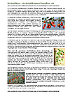
Historie und Verbreitung der Mangostan
Die ältesten Aufzeichnungen über die Nutzung der Mangostan stammen aus dem Asien des 6. Jahrhunderts n. Chr.. Um 1700 erhielt die Pflanze von dem französischen Priester und Botaniker Jacques Garcin ihren wissenschaftlichen Namen Garcinia mangostana. Der Mangostanbaum soll ursprünglich von der malaiischen Hafenstadt Malacca aus durch handelstreibende Seefahrer z. B. nach China, Indien und Ceylon (Sri Lanka) verbreitet worden sein. Englische Kolonialisten brachten die Pflanze ebenso nach Afrika, Australien, in die Karibik sowie nach Mittel- und Südamerika.
In ihren traditionellen Ursprungsgebieten wird die Pflanze auf Grund ihrer außergewöhnlichen körperstärkenden und lebensverlängernden Kräfte von den Naturvölkern seit Generationen genutzt. So werden der Baum und seine Früchte in der überlieferten Volksheilkunde bis heute bei Entzündungen, Körperschwäche, Schmerzen und verschiedenen Infektionskrankheiten, aber auch für die Schönheit innerlich und äußerlich angewendet. [7]
Mangostan-Expertin und Heilpraktikerin Karin Löprich:
„Das geheime Wissen unzähliger Generationen von Inselbewohnern aus dem asiatischen und karibischen Raum über die phänomenale Wirkung der Mangostan auf Gesundheit und Langlebigkeit ist erst durch den weltweiten Tourismus bis zu uns vorgedrungen. Kaum eine Frucht ist bei uns in der westlichen Welt so unbekannt wie die Garcinia mangostana – dabei forscht man seit vielen Jahren über sie.“ [8]
Xanthone zählen zu den wichtigsten Antioxidantien
Xanthone aus der Mangostanfrucht sind stark bioaktive Hochleistungs-Antioxidantien mit außergewöhnlichem Leistungsprofil. Antioxidantien sind Radikalfänger und damit die Gegenspieler zellschädigender und krankheitsauslösender freier Radikale. Deshalb sind Antioxidantien so essenziell wichtig für unsere Gesundheit.
Freie Radikale sind Millionen winzig kleiner, aggressiver Verbindungen, die unsere Gesundheit maßgeblich bedrohen, weil sie unsere Zellen, Gewebe und Organe schädigen, rasant altern bzw. entarten lassen, unser Immunsystem enorm schwächen und Diabetes mellitus sowie Herz-, Kreislauf-, Krebserkrankungen, Rheuma, Alzheimer, Parkinson, Grauen Star und viele weitere verheerende Erkrankungen und Folgeschäden hervorrufen bzw. beschleunigen oder verstärken können.
Die Überfrachtung des Körpers mit freien Radikalen gehört zu den gefährlichsten Gesundheitsbedrohungen unserer Zeit. Freie Radikale entstehen zwangsläufig im Stoffwechsel und zusätzlich durch unzählige äußere und körperinnere Entstehungsquellen, wie falsche Ernährung, Schadstoffe, Strahlungen, UV-Licht, Krankheitserreger, Fieber, Entzündungen, Krankheiten, emotionale Belastungen, Stress, Schlafdefizite, Gewichtsreduktion, falsch dosierter Sport u. v. a..
Unkontrolliert agierende freie Radikale sind an der Entstehung von Diabetes und anderen Radikalkrankheiten beteiligt. Die Krankheit selbst löst wiederum eine Dauerflut von freien Radikalen aus, die ihrerseits das Krankheitsgeschehen verstärken und die Selbstheilung des Körpers behindern können, da sie große Kapazitäten des Immunsystems binden.
Als typische Vertreter der Polyphenole können Xanthone in allen Körperbereichen agieren. Sie sind den Vitaminen weit überlegen. Sie gelten als Vitamin- und Antioxidantienverstärker. Das Interessante dabei: Zur „Hochform“ geraten Xanthone jedoch erst durch den Verbund mit weiteren Antioxidantien und bioaktiven Naturstoffen, denn sie besitzen bedeutende synergistische Fähigkeiten. Starke Partner für die Mangostanfrucht sind z. B. der Granatapfel, das aus roten Tomaten gewonnene Lycopin, Extrakte aus roten Traubenkernen, rote und blaue Beerenfrüchte, Acerolakirschen, Gojibeeren, Kaktusfeigen u. a.. Werden derart energiereiche Pflanzen und Naturwirkstoffe mit der Mangostanfrucht bzw. mit Xanthonen kombiniert, so führt deren Zusammenspiel automatisch zu bedeutenden gesundheitsförderlichen Ergänzungs- und Verstärkungswirkungen im Körper. Eine solche antioxidativ starke Mangostan-Fruchtkombination kann gerade den Diabetikern einen beachtlichen Gesundheitsnutzen bringen.
Diabetes ist – wie nahezu jede Krankheit - von einer chronischen Massenproduktion freier Radikale begleitet. Das wird zum Problem, wenn passgerechte Antioxidantien fehlen. Denn je schlechter das Immunsystem mangels Antioxidantien auf die Schädigungen durch freie Radikale reagieren kann, desto schneller und stärker manifestiert sich die Krankheit und desto aggressiver kann der Krankheitsverlauf sein.
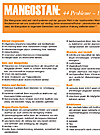
Die Mangostanfrucht kann den Insulin- und Blutzuckerhaushalt regulieren helfen
Man weiß heute, dass bei einer diabetischen Stoffwechsellage allein schon jeder Blutzuckeranstieg zu einem drastischen Anstieg von freien Radikalen in den Zellen führt. Dies hat z. B. zur Folge, dass die Zerstörung der insulinproduzierenden Betazellen der Bauchspeicheldrüse – einem zentralen Faktor der Diabeteserkrankung – beschleunigt wird. [9] Die tägliche Zufuhr ausreichender und geeigneter Antioxidantien ist die einzige Möglichkeit, dem durch radikalische Zellschäden bedingten Fortschreiten der Diabeteserkrankung entgegen zu wirken.
Hochleistungsfähige Antioxidantien wie Xanthone können die insulinproduzierenden Zellen vor Zerstörung schützen. Darüber hinaus können geeignete Antioxidantien das Insulin vor Oxidation durch freie Radikale bewahren und so die Stabilität dieses lebenswichtigen Hormons gewährleisten. Das bedeutet, dass die Wirkung des Insulins durch kraftvolle Antioxidantien verstärkt werden kann. [9] Eine verbesserte Insulinwirkung hat zur Folge, dass der Blutzuckerspiegel sinken kann bei gleichzeitiger Drosselung der Neuzuckerproduktion in der Leber. Xanthone stärken zudem jegliche Drüsenfunktionen, so auch die der Bauchspeicheldrüse und der Leber, das wurde wissenschaftlich nachgewiesen. [10], [11] Das gesunde Wechselspiel zwischen Bauchspeicheldrüse und Leber ist für uns lebenswichtig!
Bei der Blutzuckersenkung und der Verminderung von Blutzuckerschwankungen scheint auch das Pektin der Mangostanfrucht eine Rolle zu spielen. Entsprechende Wirkmechanismen beschreibt Prof. Hademar Bankhofer am Beispiel des Nopal Feigenkaktus, der pektinhaltigsten aller Pflanzen weltweit. Pektin ist ein löslicher Ballaststoff, der die Fähigkeit hat, einen Teil der aus der Nahrung stammenden Glucose (Zucker) zu binden, diese vorzeitig zur Ausscheidung zu bringen und sie so am Verdauungsgeschehen, also auch am Eintritt in die Blutgefäße vorbei zu schleusen. [12]
Der Blutzucker kann außerdem durch die Blockierung von stärkeabbauenden Verdauungsenzymen im Darm gesenkt werden. Dadurch wird Stärke viel langsamer zu Glucose (Zucker) umgewandelt. Daraus resultiert eine zeitverzögerte Abgabe der Glucose an das Blut. So kommt es zu einem langsameren und reduzierten Anstieg des Zuckerspiegels im Blut. [6]
Eine Verbesserung des Blutzuckerhaushaltes kann sich in gesunkenen Blutzucker-Nüchternwerten oder im niedrigeren Blutzucker-Langzeitwert (HbA1-c) niederschlagen.
Radikalfangende Polyphenole aus der Mangostan können Entzündungen reduzieren
Eine Folge des Diabetes ist grundsätzlich ein mehr oder weniger ausgeprägtes Entzündungsgeschehen. Daraus folgt wiederum zwangsläufig ein chronisches Übermaß an zellschädigenden freien Radikalen. Diese „nähren“ die Entzündungsprozesse, wenn Antioxidantien fehlen. Wissenschaftler haben nachgewiesen, dass phenolische Xanthone nicht nur außergewöhnliche antioxidative Fähigkeiten haben, sondern auch hervorragende entzündungshemmende Eigenschaften besitzen. Besonders bei Diabetes kann deshalb ein hochleistungsfähiger synergistischer Antioxidantienverbund auf Xanthonebasis das hoffnungslos überforderte Immunsystem spürbar, sichtbar und messbar entlasten: Beispielsweise kann sich die Verbesserung des Antioxidantien-Spiegels im Körper in beschleunigter Wundheilung, Schmerzreduzierung und Schwellungsrückgang, aber auch in geringerer Infektanfälligkeit bzw. in verbessertem Allgemeinbefinden auswirken. Zu messen ist der entzündungshemmende Einfluss von Antioxidantien beispielsweise am reduzierten Blutsenkungswert.
Polyphenole bzw. Xanthone sind in der Lage, die fetthaltigen und deshalb besonders empfindlichen Nervenzellstrukturen vor Radikalschädigungen und Entzündungen zu bewahren. Davon kann letztlich das gesamte Nervensystem einschließlich Gehirn profitieren.
Xanthone können die Durchblutung fördern
Des Weiteren sind Polyphenole, zu denen die Xanthone zählen, bekannt für ihre blutgefäßschützenden und blutdruckregulierenden Eigenschaften. Insbesondere die mikrokapillare Blutzirkulation – und damit auch die gefährdeten Augen- und Nierenfunktionen - profitieren von den durchblutungsfördernden Merkmalen der Polyphenole.
Polyphenole bzw. Xanthone wirken Arteriosklerose und Thrombose entgegen, das wurde wissenschaftlich nachgewiesen. Polyphenole können die Zusammensetzung der Blutfette verbessern und den Cholesterin-Haushalt regulieren: Sie sind in der Lage, die Oxidation von LDL-Cholesterin im Blut zu verhindern. Das haben australische Forscher herausgefunden. Die Beweglichkeit der roten Blutkörperchen wird durch den Einfluss von Polyphenolen erhöht, das Blut wird fließfähiger, die Gefäße erhalten eine höhere Elastizität, die Durchblutungsrate und die damit verbundene Sauerstoffversorgung steigen. [11], [13] Dies ist von entscheidender Bedeutung z. B. für die stark gefährdeten peripheren Nerven (Nerven der Gliedmaßen) von Diabetikern. Die verbesserte Durchblutung kann beispielsweise am regulierten Blutdruck gemessen werden.
Mangostan-Früchteverbund in flüssiger Form
Wahrscheinlich sind die begrenzten, sehr fern gelegenen und stark klimaabhängigen Anbauregionen von Mangostanbäumen, verbunden mit der kurzen Haltbarkeit der Früchte, wesentliche Gründe, warum sie in unseren Breiten so lange unbekannt geblieben sind. Die Chance, hierzulande Qualitätsfrüchte zu erstehen, sind auf Grund des langen Transportweges leider nur gering. Einmal geerntete Mangostanfrüchte reifen nicht mehr nach, werden schnell hart und verderben schon nach wenigen Tagen. Auch kleinste Haarrisse in der Schale durch leichte Schädigungen bei Lagerung und Transport lassen das empfindliche weiße Fruchtinnere schnell verderben.
Die empfehlenswerte Alternative: Mangostan in flüssiger Form!
Mit Blickpunkt auf einen höchstmöglichen antioxidativen, antientzündlichen und durchblutungsfördernden Wirkungsgrad ist für den Typ 1- und Typ 2-Diabetiker eine Kombination aus der xanthonereichen Mangostanfrucht und weiteren sinnvollen antioxidantien- und bioaktivstoffreichen Pflanzen und Früchten unter Einbeziehung von Wirkstoffaufkonzentrierungen als flüssige Darreichungsform anzuraten. Die Begründung ist einfach: Die Mangostan besitzt ausgeprägte synergistische Eigenschaften, die gerade bei Diabetes für gesundheitsunterstützende Maximaleffekte voll ausgeschöpft werden sollten.
Geachtet werden muss hierbei auf den Verzicht sowohl von Zucker- und Süßstoffzusätzen als auch von synthetischen Farb-, Aroma- und Konservierungsstoffen.
Verfasst & Copyright:
Katrin Nehls
Diplom-Volkswirtin
Unabhängige, freie Medizin- und Gesundheitsredakteurin
Verfasst im Auftrag des „Instituts für
Mangostan & natürlichen Antioxidantien“
www.Mangostan-Institut.com
Quellen:
[1] Subrt, I.: Erfahrungsbericht, Mangostan-Gold Ltd. & Co KG, Berlin 2008
[2] Beck, R.: Erfahrungsbericht, Mangostan-Gold Ltd. & Co KG, Berlin 2008
[3] Röll, R.: Erfahrungsbericht, Mangostan-Gold Ltd. & Co KG, Berlin 2008
[4] Dr. Müller-Wohlfahrt, H.-W.: „So schützen Sie Ihre Gesundheit“ Deutscher Taschenbuchverlag, München 2005
[5] Prof. Hoffmann, M. et al.: „Vom Lebendigen in Lebensmitteln“ Stiftung
Ökologie & Landbau (SÖL), Deukalion Verlag, Holm 1997
[6] Dr. Watzl, B., Prof. Leitzmann, C.: „Bioaktive Substanzen in Lebensmitteln“ 3. unveränd. Aufl., Hippokrates Verlag, Stuttgart 2005
[7] Reiseerzählungen. Interview mit Rolf Hausmann am 22.01.2008
[8] Löprich, K.: „Naturheilmittel Mangosteen: Königin der Früchte!“
http://www.chelat.biz/DATEN/neu/ETDA-Beitrag_MH_2006.pdf
[9] Dr. Schelosky, S. „Diabetes mellitus und Neurodegeneration: Freie Radikale eine Ursache“, Pressemitteilung, Deutsches Institut für Ernährungsforschung Potsdam-Rehbrücke, 15.08.2003
[10] Dr. med. Huber,R.: “Die Mangostanfrucht auf dem Weg zum natürlichen Super-Antioxidant?” Institut für Mangostan und natürliche Antioxidantien, 2007
[11] Simonsohn, B.: „Heilkraft aus den Tropen. Die süße Medizin exotischer Früchte“ Integral-Verlag, 2008
[12] Prof. Bankhofer, H. et al.: „Die heilenden Kräfte des Nopal“ Kneipp Verlag, 2. Aufl., Leoben 2004
[13] Zeitschrift für Phytotherapie 5/2007, 28: 220, Hippokrates Verlag
Prof. Ohlenschläger, G.: „Freie Radikale, Oxidativer Stress und Antioxidantien“ 2. erw. Aufl., Ralf Reglin Verlag, Köln 2000
Dr. Huber, R., Dr. Ranft, R.: „mangostan“ Verlag Carl Ueberreuter, Wien 2008
Nehls, K.: „ Kraftvoller Zellschutz gegen freie Radikale mit der Mangostan“
1. Aufl., Institut für Mangostan und natürliche Antioxidantien, 2008
Nehls, K.: „Mit der Kraft der Mangostan. Flüssiges Multitalent bekämpft Entzündungen und stärkt das Immunsystem“, Natur-Heilkunde Journal. Medizin Praxis Wissenschaft, Sonderdruck. 04/2008
Diabetes
Aktuelle wissenschaftliche Studien
Quelle: Datenbank der U.S. National Library of Medicine and the National Institutes of Health (siehe http://www.ncbi.nlm.nih.gov/sites/entrez)
1: Pancreas. 2007 Nov;35(4):e10-7.
Related Articles,
Links
![]()
Reduction of oxidative stress by a new low-molecular-weight antioxidant improves metabolic alterations in a nonobese mouse diabetes model.
Novelli M, D'Aleo V, Lupi R, Paolini M, Soleti A, Marchetti P, Masiello P.
Dipartimento di Patologia Sperimentale, University of Pisa, Pisa, Italy.
OBJECTIVES: We have previously established a nonobese diabetes mouse model characterized by moderate hyperglycemic levels, like those usually occurring in human type 2 diabetes. As oxidative stress is considered a major mechanism of progressive beta-cell damage in diabetes, we tested in this model the protective effects of a new low-molecular-weight antioxidant, namely, bis(1-hydroxy-2,2,6,6-tetramethyl-4-piperidinyl)decandioate dihydrochloride (IAC). METHODS: Diabetes was induced in C57Bl/6J mice by streptozotocin (STZ) and nicotinamide (NA) administration. Two weeks later, STZ-NA mice were treated for 5 weeks with different doses of IAC (15 or 30 mg/kg per day intraperitoneally) and monitored for glycemia, insulinemia, glucose tolerance, and pancreatic insulin content. RESULTS: Streptozotocin-NA mice showed moderate hyperglycemia, hypoinsulinemia, glucose intolerance, growth impairment, and markedly reduced pancreatic insulin content (22% of controls). IAC-treated STZ-NA mice showed clear-cut reduction of hyperglycemia and attenuation of glucose intolerance, associated to higher residual pancreatic insulin content with respect to untreated diabetic animals. Plasma nitrotyrosine levels (an index of oxidative stress), enhanced 3-fold in diabetic mice, were significantly reduced by IAC treatment. Significant correlations were found between plasma nitrotyrosine values and either blood glucose levels or pancreatic insulin content. CONCLUSIONS: In the STZ-NA diabetic mouse model, the new antioxidant, IAC, improves diabetic metabolic alterations, likely by counteracting beta-cell dysfunction and loss associated with oxidative stress.
Publication Types:
PMID: 18090226 [PubMed - indexed for MEDLINE]
2: Diabetes Res Clin Pract. 2007 Sep;77(3):427-32. Epub 2007 Mar 13.
Related Articles,
Links
![]()
Erythrocyte susceptibility to oxidative stress and antioxidant status in patients with type 1 diabetes.
Firoozrai M, Nourbakhsh M, Razzaghy-Azar M.
Department of Biochemistry, School of Medicine, Iran University of Medical Sciences, Tehran, Iran.
In this study, malondialdehyde (MDA) level as an index of erythrocyte susceptibility to oxidative stress and antioxidant defense system (glutathione level (GSH), glutathione peroxidase enzyme activity (GPx) in erythrocytes and ferric reducing ability of plasma (FRAP) as the total plasma antioxidant capacity were measured in 35 patients with type 1 diabetes and 28 age and sex-matched normal subjects. MDA level was significantly elevated in diabetic patients (650.9+/-144.3 nmol/g versus 476.5+/-138.5 nmol/g Hb, P<0.001). The level of MDA was positively correlated with duration of diabetes (r= 0.29, P<0.05) and HbA(1C) (r= 0.39, P<0.05) and negatively with FRAP (r= -0.3, P<0.05). The level of GSH and FRAP were lower in patients than controls (7.05+/-1.6 micromol/g versus 8.24+/-0.9 micromol/g Hb, and 389.05+/-82.3 micromol/l versus 520.4+/-124.1 micromol/l, respectively, P<0.001). GPx activity was not significantly different between the two groups. GSH and FRAP were negatively correlated with HbA(1C) (r= -0.334, P<0.01 and r= -0.5, P<0.01, respectively). In conclusion, there seems to be an increased susceptibility to oxidative stress and decreased antioxidant defense in patients with type 1 diabetes, which may be due to poor glycemic control.
PMID: 17360068 [PubMed - indexed for MEDLINE]
3: Clin Sci (Lond). 2007 Jun;112(12):599-606.
Related Articles,
Links
![]()
Oxidative stress, antioxidant status and DNA damage in patients with impaired glucose regulation and newly diagnosed Type 2 diabetes.
Song F, Jia W, Yao Y, Hu Y, Lei L, Lin J, Sun X, Liu L.
Department of Nutrition and Food Hygiene, School of Public Health, Tongji Medical College, Huazhong University of Science and Technology, Wuhan, People's Republic of China.
Previous studies have postulated the association between oxidative stress and Type 2 diabetes. Considering the long pre-diabetic period with IGR (impaired glucose regulation) and its high risk of developing diabetes, to test this hypothesis, we have investigated oxidative stress pathways and DNA damage in patients with IGR and newly diagnosed Type 2 diabetes. The study population consisted of 92 subjects with NGT (normal glucose tolerance), 78 patients with IGR and 113 patients with newly diagnosed diabetes. Plasma MDA (malondialdehyde) and TAC (total antioxidative capacity) status, erythrocyte GSH content and SOD (superoxide dismutase) activity were determined. A comet assay was employed to evaluate DNA damage. Compared with subjects with NGT, patients with IGR had reduced erythrocyte SOD activity. Patients with diabetes had a higher plasma MDA concentration, but a lower plasma TAC level and erythrocyte SOD activity, than the NGT group. Correlation analysis revealed a strong positive association between IR (insulin resistance) and MDA concentration, but negative correlations with TAC status and SOD activity. With respect to beta-cell function, a positive association with TAC status and an inverse correlation with GSH respectively, were observed. The comet assay revealed slight DNA damage in patients with IGR, which was increased in patients with diabetes. Significant correlations were observed between DNA damage and hyperglycaemia, IR and beta-cell dysfunction. In conclusion, the results of the present study suggest that hyperglycaemia in an IGR state caused the predominance of oxidative stress over antioxidative defence systems, leading to oxidative DNA damage, which possibly contributed to pancreatic beta-cell dysfunction, IR and more pronounced hyperglycaemia. This vicious circle finally induced the deterioration to diabetes.
Publication Types:
PMID: 17209802 [PubMed - indexed for MEDLINE]
4: Clin Ther. 2005 Nov;27(11):1764-73.
Related Articles,
Links
![]()
Effects of antioxidant supplementation on postprandial oxidative stress and endothelial dysfunction: a single-blind, 15-day clinical trial in patients with untreated type 2 diabetes, subjects with impaired glucose tolerance, and healthy controls.
Neri S, Signorelli SS, Torrisi B, Pulvirenti D, Mauceri B, Abate G, Ignaccolo L, Bordonaro F, Cilio D, Calvagno S, Leotta C.
Department of Internal Medicine and Systemic Disease, University of Catania, Via S. Sofia 86, 95123 Catania, Italy. sergio.neri4@tin.it
BACKGROUND: Increased generation of reactive oxygen species (ROS) and oxidative stress may be of crucial importance in the pathogenesis of endothelial damage. Furthermore, there is understood to be a relationship between endothelial damage, glycemic control, disorders of lipid metabolism, and coagulative hemostatic disorders. OBJECTIVE: This study investigated within- and between-group changes in various circulating markers of oxidation-reduction balance and endothelial function after a balanced moderate-fat meal with and without antioxidant supplementation in patients with early-stage, untreated type 2 diabetes mellitus; subjects with impaired glucose tolerance (IGT); and healthy controls. METHODS: In this single-blind, controlled clinical study, groups of patients with type 2 diabetes and subjects with IGT were identified and compared with a group of healthy controls. All groups followed a controlled, well-balanced diet for 10 days before and throughout the study. Before and after consumption of a standardized moderate-fat meal, plasma levels of oxidants (malondialdehyde, 4-hydroxynonenal, oxidized low-density lipoprotein), the antioxidant glutathione peroxidase, and markers of endothelial function (NO, endothelin-1, von Willebrand factor [vWF], vascular cell adhesion molecule-1 [VCAM-1]) were determined. These measures were then reassessed after 15 days of standard antioxidant treatment consisting of a thiol-containing antioxidant (N-acetylcysteine 600 g/d), a bound antioxidant (vitamin E 300 g/d), and an aqueous phase antioxidant (vitamin C 250 mg/d). The efficacy of antioxidant treatment in reversing abnormalities in oxidation-reduction balance after a moderate-fat meal was assessed by evaluating changes in plasma levels of ROS on the morning of the 16th day following an overnight fast. Safety was monitored in terms of adverse events, vital signs, physical findings, and laboratory values. RESULTS: The study included 46 patients with type 2 diabetes (23 men, 23 women; mean [SD] age, 41 [3] years; mean body mass index [BMI], 24 [2] kg/m(2)), 46 with IGT (23 men, 23 women; mean age, 39 [3] years; mean BMI, 23 [3] kg/m(2)), and 46 control subjects (23 men, 23 women; mean age, 40 [1] years; mean BMI, 22 [1] kg/m(2)). Before supplementation, all 3 groups had significantly increased levels of oxidants, vWF, and VCAM-1 (all, P < 0.001) and significantly decreased levels of antioxidants and NO (both, P < 0.001) after consumption of a moderate-fat meal. After 15 days of antioxidant treatment, significant improvements in these measures were seen in all groups (P < 0.05). CONCLUSIONS: This study showed changes in oxidation-reduction balance, NO bioavailability, and nonthrombogenic endothelial factors after a moderate-fat meal in patients with type 2 diabetes and those with IGT, but these postprandial changes were reverse in all subjects after 15 days of standard antioxidant supplementation. These findings suggest that the use of anti-oxidants may have decreased oxidative stress in these subjects.
Publication Types:
PMID: 16368447 [PubMed - indexed for MEDLINE]
5: Ann N Y Acad Sci. 2004 Dec;1031:204-13.
Related Articles,
Links
![]()
Oxidative stress and antioxidant treatment in diabetes.
Scott JA, King GL.
Research Division, Joslin Diabetes Center, Harvard Medical School, One Joslin Place, Boston, MA 02215, USA.
The many studies on oxidative stress, antioxidant treatment, and diabetic complications have shown that oxidative stress is increased and may accelerate the development of complications through the metabolism of excessive glucose and free fatty acids in diabetic and insulin-resistant states. However, the contribution of oxidative stress to diabetic complications may be tissue-specific, especially for microvascular disease that occurs only in diabetic patients but not in individuals with insulin resistance without diabetes, even though both groups suffer from oxidative stress. Although antioxidant treatments can show benefits in animal models of diabetes, negative evidence from large clinical trials suggests that new and more powerful antioxidants need to be studied to demonstrate whether antioxidants can be effective in treating complications. Furthermore, it appears that oxidative stress is only one factor contributing to diabetic complications; thus, antioxidant treatment would most likely be more effective if it were coupled with other treatments for diabetic complications.
Publication Types:
PMID: 15753146 [PubMed - indexed for MEDLINE]
6: Arch Gerontol Geriatr. 2004 Nov-Dec;39(3):269-75.
Related Articles,
Links
![]()
Oxidative stress and antioxidant status in elderly diabetes mellitus and glucose intolerance patients.
Atli T, Keven K, Avci A, Kutlay S, Turkcapar N, Varli M, Aras S, Ertug E, Canbolat O.
Department of Geriatric Medicine, Ankara University School of Medicine, Cebeci, 06110, Turkey. teslimeatli@yahoo.com
Increased oxidative stress and impaired anti-oxidant defense have been suggested as contributory factors for initiation and progression of complications in diabetes mellitus. Aging itself has been shown to be along with increased oxidative stress and lower anti-oxidant defense. We aimed at investigating oxidative stress and anti-oxidant enzymes in 61 elderly subjects. Fifteen healthy individuals (group 1, mean age 72.2 +/- 5.13), 13 glucose intolerant patients (group 2, mean age 71.7 +/- 4.9), 19 patients with type 2 diabetes mellitus (T2DM) without any complication (group 3, mean age 70.0 +/- 6.0), and 14 patients with T2DM with at least one complication (group 4, mean age 69.8 +/- 4.7) were included in the study. Whilst plasma levels for malondialdehyde (MDAP) and erythrocyte malondialdehyde (MDAE) were measured as markers of oxidative stress, activity of erythrocyte superoxide dismutase (SOD), glutathion peroxidase (GSH-Px), and catalase (CAT) were taken as markers of oxidative defense system. MDAP level was significantly elevated in group 4 (P = 0.001). MDAE was elevated in patients with T2DM, particularly in group 4, however, the difference between the groups was of borderline significance (P = 0.07). Whilst CAT was elevated in groups 3 and 4 compared to control subjects (P = 0.025 and 0.002, respectively), no difference was found for SOD between the groups. GSH-Px activity was found to be increased in groups 2, 3 and 4, it did not reach statistical significance (P = 0.106). There were significant correlations between CAT and MDAE (P < 0.0001, r = 0.056) and MDAP (P = 0.016, r = 0.306). These results suggest that there was an increased oxidative stress in elderly diabetics, however, this is not due to reduced erythrocyte antioxidant defense potential but, rather, increased free radical production possibly due to hyperglycemia.
PMID: 15381345 [PubMed - indexed for MEDLINE]
7: J Androl. 2004 Sep-Oct;25(5):830-6.
Related Articles,
Links
![]()
Oxidative stress and antioxidant therapy: their impact in diabetes-associated erectile dysfunction.
De Young L, Yu D, Bateman RM, Brock GB.
Department of Urology, St Joseph's Health Care, London, Canada N6A 4V2.
Oxidative stress is believed to affect the development of diabetic-associated vasculopathy, endothelial dysfunction, and neuropathy within erectile tissue. Our hypothesis is that, given adequate concentrations of the oxygen free radical scavenger vitamin E, enhanced levels of circulating nitric oxide (NO) should improve erectile function with the potential for a synergistic effect with a phosphodiesterase type 5 (PDE5) inhibitor. Twenty adult male Sprague-Dawley streptozotocin-induced (60 mg/kg intraperitoneally) diabetic rats were placed in 4 therapeutic groups (n = 5 per group) as follows: 1) peanut oil only (diabetic control), 2) 20 IU of vitamin E per day, 3) 5 mg/kg of sildenafil per day, and 4) vitamin E plus sildenafil using oral gavage for 3 weeks. In addition, 5 age-matched rats served as normal nondiabetic controls (normal). Erectile function was assessed by measuring the rise in intracavernous pressure (ICP) following cavernous nerve electrostimulation. Penile tissue was evaluated for neuronal NO synthase (nNOS), smooth muscle alpha-actin, nitrotyrosine, and endothelial cell integrity. Urine nitrite and nitrate (NOx) concentration was quantified, and electrolytes were tested by a serum biochemistry panel. A significant decrease in ICP was recorded in the diabetic animals, with improvement measured in the animals receiving PDE5 inhibitors either with or without vitamin E; the controls had a pressure of 54.8 +/- 5.3 cm H2O, the vitamin E group had a pressure of 73.5 +/- 6.6 cm H2O, the sildenafil group had a pressure of 78.4 +/- 10.77 cm H2O, and the vitamin E plus sildenafil group had a pressure of 87.9 +/- 5.5 cm H2O (P <.05), compared with the normal cohorts at 103.0 +/- 4.8 cm H2O. Histoexaminations showed improved nNOS, endothelial cell, and smooth muscle cell staining in the vitamin E plus sildenafil group compared to the control animals. Urine NOx increased significantly in all the diabetic groups but was blunted in the vitamin E and vitamin E plus sildenafil groups. A significant increase in positive staining for nitrotyrosine was observed in the vitamin E plus sildenafil group. Vitamin E enhanced the therapeutic effect of the PDE5 inhibitor in this study, supporting the potential use of oxygen free radical scavengers in salvaging erectile function in diabetic patients.
Publication Types:
PMID: 15292117 [PubMed - indexed for MEDLINE]
8: J Investig Med. 2004 Jan;52(1):19-23.
Related Articles,
Links
Role of oxidative stress in the etiology of type 2 diabetes and the effect of antioxidant supplementation on glycemic control.
Opara EC.
Department of Surgery, Duke University Medical Center, Durham, NC, USA. opara001@hotmail.com
Oxidative stress is a situation in which the amount of reactive oxygen species (ROS) exceeds the levels of neutralizing substances referred to as antioxidants. Numerous studies have shown that oxidative stress is associated with type 2 diabetes, and there is compelling biochemical evidence that suggests that ROS may even play a role, if only secondary, in the pathogenesis of type 2 diabetes. These observations have provided sufficient impetus for the use of antioxidant supplements as adjunct therapy for control of blood sugar in diabetic patients. However, there is currently no optimum regimen of antioxidant supplementation for diabetic patients. Studies are required to determine appropriate doses of relevant individual micronutrients that perhaps should be used in combination to diminish oxidative stress and improve glycemic control in individuals afflicted with type 2 diabetes.
Publication Types:
PMID: 14989366 [PubMed - indexed for MEDLINE]
9: Front Biosci. 2004 Jan 1;9:565-74.
Related Articles,
Links
![]()
Mechanisms of oxidative stress in diabetes: implications for the pathogenesis of vascular disease and antioxidant therapy.
Pennathur S, Heinecke JW.
Department of Medicine, Division of Metabolism, Endocrinology, and Nutrition, University of Washington, Seattle WA 98195, USA. spenath@u.washington.edu
Diabetes markedly raises the risk of microvascular and macrovascular disease, the major contributors to higher morbidity and mortality in this increasingly prevalent disorder. Oxidative stress has been postulated as one major contributor to long-term diabetic complications. However, there is considerable controversy regarding the nature, magnitude, and mechanisms of oxidative stress in the diabetic state. Although products of glycoxidation and lipoxidation are elevated in plasma and tissue from humans suffering from diabetes, the exact relationships among hyperglycemia, the diabetic state, and oxidative stress are undetermined. This review focuses on proposed mechanisms for increasing oxidative stress in diabetes, the relationship of oxidant production to hyperglycemia, the contribution of reactive carbonyl compounds that accumulate in the diabetic state to tissue injury, and the potential role of lipids in producing oxidants. Current evidence argues against a generalized increase in oxidative stress in human diabetes, at least in the extracellular milieu. Instead, reactive intermediates generated in certain microenvironments might promote oxidative stress by unique pathways. Thus, many issues need to be addressed, including the suitability of antioxidants for preventing the clinical sequelae of diabetes.
Publication Types:
PMID: 14766391 [PubMed - indexed for MEDLINE]
10: J Neurol Sci. 2004 Mar 15;218(1-2):17-24.
Related Articles,
Links
![]()
Oxidative stress in Alzheimer's and vascular dementias: masking of the antioxidant profiles by a concomitant Type II diabetes mellitus condition.
Serra JA, Marschoff ER, Domínguez RO, Guareschi EM, Famulari AL, Pagano MA, de Lustig ES; Collaborative Group for the Study of the Oxidative Stress, Argentina.
Collaborative Group for the Study of the Oxidative Stress, Argentina.
Oxidative stress is associated with Alzheimer's (DAT) and vascular (VD) dementias, as well as Type II diabetes mellitus (DIAB) and affected by hypoglycemic therapy. The population (n = 122; males = 60; mean age = 72.57 +/- 7.06) consisted of controls (CTR), DAT and VD patients, with (DAT + DIAB, VD + DIAB) and without concomitant DIAB, resulting in six groups where the antioxidant profile was determined: copper-zinc superoxide dismutase (SOD), thiobarbituric acid reactive substances (TBARS), and total antioxidant capacity (TRAP). The results were analyzed using a two-way ANOVA design and Bonferroni statistic. The ANOVAs yielded significant differences between groups for all components of the profile: SOD, p = 0.00000006; TBARS, p = 0.0000012; TRAP, p = 0.0000003. The significance level for comparisons between groups was set at alpha = 0.05. The comparisons DIAB vs. CTR, DAT+DIAB vs. DAT, and DIAB demented vs. DIAB non-demented resulted significant for all variables. VD + DIAB vs. VD resulted significant for all variables except TRAP. The antioxidant profiles of DIAB and CTR are different. The differences cannot be directly related with what is observed in dementias. The differences in profiles of demented and non-demented are somewhat hidden when demented patients are affected by a concomitant DIAB condition and/or hypoglycemic treatment, thus conditioning the diagnostic value for dementias of the profiles.
Publication Types:
PMID: 14759628 [PubMed - indexed for MEDLINE]
11: Free Radic Biol Med. 2003 Jun 15;34(12):1563-74.
Related Articles,
Links
![]()
Biomarkers of diabetes-associated oxidative stress and antioxidant status in young diabetic patients with or without subclinical complications.
Martín-Gallán P, Carrascosa A, Gussinyé M, Domínguez C.
Biochemistry and Molecular Biology Center, Hospital Universitario Vall d'Hebron, Barcelona, Spain.
The aims of the study were to ascertain the potential role of oxidative stress in the onset of disease-related pathophysiological complications in young type 1 diabetes patients. Indicative parameters of lipoperoxidation, protein oxidation, and changes in antioxidant defense system status were measured in blood samples from 26 young diabetic patients with recently diagnosed (< 6 months) microangiopathy (+DC), 28 diabetic patients without complications (-DC), and 40 healthy age-matched controls (CR). Both diabetic groups presented similar fructosamine and glycated hemoglobin (HbA1c) values. Results showed erythrocyte glutathione peroxidase activity, glutathione content, and plasma beta-carotene to be significantly lower in diabetic patients compared with control subjects, but with no significant differences between -DC and +DC groups. Antioxidant enzyme superoxide dismutase activity was significantly higher in the erythrocytes of diabetic patients independently of the presence of microvascular complications. However, the plasma alpha-tocopherol/total lipids ratio was significantly diminished in +DC group compared with -DC (p =.008). Lipid peroxidation indices measured in plasma included malondialdehyde, lipid hydroperoxides, and lipoperoxides, which were significantly elevated in our diabetic patients regardless of the presence of complications. Evidence of oxidative damage to proteins was shown both through the quantification of plasma protein carbonyl levels, which were significantly higher in -DC (0.61 +/- 0.09 mmol/mg prot), and higher still in the +DC patients (0.75 +/- 0.09 mmol/mg prot) compared with those of controls (0.32 +/- 0.03 mmol/mg prot; p <.01) and immunoblot analysis of protein-bound carbonyls. Additionally, a marked increase in protein oxidation was observed in +DC patients through assessment of advanced oxidation protein products (AOPP) considered to be an oxidized albumin index; AOPP values were significantly higher in +DC than in -DC patients (p <.01) and CR (p <.0001). These results point to oxidatively modified proteins as a differential factor possibly related to the pathogenesis of diabetic complications.
Publication Types:
PMID: 12788476 [PubMed - indexed for MEDLINE]
12: Diabetes Obes Metab. 2002 Sep;4(5):305-8.
Related Articles,
Links
![]()
Relationship of glycation, antioxidant status and oxidative stress to vascular endothelial damage in diabetes.
Wen Y, Skidmore JC, Porter-Turner MM, Rea CA, Khokher MA, Singh BM.
School of Health Sciences, University of Wolverhampton, Wolverhampton, UK.
AIMS: To examine the inter-relationships of various microvascular pathogenic mechanisms in diabetic patients. METHODS: Patients with diabetes (n = 18) and non-diabetic subjects (n = 18) were studied. RESULTS: Blood markers of glycaemic control and glycation differed between the two groups (glucose 10.9 +/- 7.6 vs. 4.7 +/- 0.63 mmol/l, p < 0.01; HbA1c 7.0 +/- 1.3 vs. 4.5 +/- 0.3%, p < 0.001; glycated LDL 8.8 +/- 2.5 vs. 6.1 +/- 1.2%, p < 0.001) but plasma antioxidant status did not. LDL oxidation resistance, measured as lag time to maximum oxidation initiated by copper ions, was decreased in diabetes (58. +/- 14.3 vs. 76.3 +/- 21.5 min, p < 0.01). Both soluble intercellular adhesion molecule-1 (ICAM-1) and vascular cell adhesion molecule-1 (VCAM-1), markers of endothelial dysfunction, were significantly higher in diabetes (ICAM 491 +/- 128 vs. 403 +/- 131 micro g/l, p < 0.05; VCAM 546 +/- 157 vs. 393 +/- 106 micro g/l, p < 0.01). Linear correlations were significant between HbA1c and lag time of LDL oxidation (r = -0.39, p < 0.05), ICAM (r = 0.40, p < 0.05) and VCAM (r = 0.38, p < 0.05). LDL oxidizability correlated with vitamin C (r = 0.51, p < 0.01) but not any adhesion molecule. In multivariate analysis, both ICAM and VCAM correlated with HbA1c only (r(2) = 0.16, F = 6.3, p < 0.01; r(2) = 0.14, F = 5.4, p < 0.01 respectively). CONCLUSION: In diabetes, glycation, tissue oxidation and endothelial function are all abnormal and predisposing to microvascular complications but interrelationships are complex with glycation appearing most direct.
PMID: 12190993 [PubMed - indexed for MEDLINE]
13: J Intern Med. 2002 Jan;251(1):69-76.
Related Articles,
Links
![]()
Oxidative stress and antioxidant status in type 1 diabetes mellitus.
Vessby J, Basu S, Mohsen R, Berne C, Vessby B.
Section for Clinical Nutrition Research, Department of Public and Caring Sciences, Uppsala University, Uppsala, Sweden. johanvessby@hotmail.com
OBJECTIVES: To test the hypothesis that type 1 diabetes is associated with increased oxidative stress and/or antioxidant status by investigating concentrations of 8-iso-prostaglandin F2alpha (8-iso-PGF2alpha) in urine and plasma and malondialdehyde (MDA) in plasma as indicators of lipid peroxidation in vivo, and antioxidant status in diabetic subjects compared with healthy control subjects. DESIGN AND SUBJECTS: Thirty-eight subjects with type 1 diabetes mellitus and 41 healthy age- and sex-matched control subjects were included in the study. Blood and urine samples were obtained and analysed for 8-iso-PGF2alpha with a newly developed radioimmunoassay, as well as for MDA, total antioxidant capacity (TAOC) and serum tocopherol levels. RESULTS: None of the variables of lipid peroxidation showed any significant difference between the two groups. Similarly, there were no significant correlations between the levels of 8-iso-PGF2alpha or MDA, and degree of glycemic control (HbA1c). Total antioxidant capacity in plasma was 16% lower amongst the subjects with type 1 diabetes than in the control group (P < 0.0005). Lipid corrected levels of alpha-tocopherol in serum were significantly increased in type 1 diabetic subjects (P < 0.05), as were gamma-tocopherol levels (P < 0.005). CONCLUSIONS: In spite of lower total antioxidant defence, our results do not support the oxidative stress hypothesis for type 1 diabetes mellitus. The higher tocopherol levels suggest that no vitamin E supplementation is necessary for subjects with type 1 diabetes mellitus.
Publication Types:
PMID: 11851867 [PubMed - indexed for MEDLINE]
14: Diabetes Care. 2002 Feb;25(2):370-5.
Related Articles,
Links
![]()
Early increase of oxidative stress and reduced antioxidant defenses in patients with uncomplicated type 1 diabetes: a case for gender difference.
Marra G, Cotroneo P, Pitocco D, Manto A, Di Leo MA, Ruotolo V, Caputo S, Giardina B, Ghirlanda G, Santini SA.
Institute of Internal Medicine and Geriatrics, Catholic University School of Medicine, Largo Gemelli 8, 00168 Rome, Italy.
OBJECTIVE: Diabetes increases the risk of coronary heart disease (CHD) to a greater extent in women than in men. We investigated whether type 1 diabetic patients with short duration of disease and without complications have an altered oxidative status and whether there are differences between men and women. RESEARCH DESIGN AND METHODS: We investigated oxidative status in 29 control subjects and 37 patients with uncomplicated type 1 diabetes with duration of 6 +/- 3 years. RESULTS: Compared with control subjects, type 1 diabetic patients had lower total plasma antioxidant capacity (TRAP) (720.3 +/- 111.2 vs. 972.5 +/- 97.7 micromol/l in men, P < 0.001; 579.8 +/- 95.4 vs. 930.1 +/- 84.2 in women, P < 0.001), higher lipid hydroperoxide (ROOH) levels (6.4 +/- 2.2 vs. 2.0 +/- 0.7 micromol/l in men, P < 0.001; 8.1 +/- 1.9 vs. 2.2 +/- 0.6 in women, P < 0.001), higher total conjugated diene (CD) levels (0.037 +/- 0.003 vs. 0.033 +/- 0.002 A.U. in men, P < 0.001), lower 246-nm CD levels (0.0032. +/- 0.0010 vs. 0.0070 +/- 0.0012 A.U. in men, P < 0.001; 0.0022 +/- 0.0011 vs. 0.0072 +/- 0.0014 A.U. in women, P < 0.001), and higher 232-nm CD levels (0.0348 +/- 0.0041 vs. 0.0257 +/- 0.0022 A.U. in men, P < 0.001; 0.0346 +/- 0.0031 vs. 0.0246 +/- 0.0074 A.U. in women, P < 0.001). Compared with diabetic men, diabetic women had lower TRAP (P < 0.01), higher ROOH levels (P < 0.01), and lower 246-nm CD levels (P < 0.05). Plasma concentration of uric acid was significantly lower in patients with type 1 diabetes than in control subjects (3.3 +/- 0.3 vs. 4.3 +/- 0.2 mg/dl; P = 0.009) with a significant difference between women and men with type 1 diabetes (2.6 +/- 0.3 vs. 3.9 +/- 0.3, respectively; P = 0.009). CONCLUSIONS: Our findings suggest that reduced antioxidant activity and increased oxidative stress occur early after the diagnosis of type 1 diabetes, especially in women, and this might explain, at least in part, the increased susceptibility of diabetic women to cardiovascular complications.
Publication Types:
PMID: 11815512 [PubMed - indexed for MEDLINE]
15: Diabetologia. 2000 Aug;43(8):974-7.
Related Articles,
Links
![]()
Oral treatment with an antioxidant (raxofelast) reduces oxidative stress and improves endothelial function in men with type II diabetes.
Chowienczyk PJ, Brett SE, Gopaul NK, Meeking D, Marchetti M, Russell-Jones DL, Anggård EE, Ritter JM.
Department of Clinical Pharmacology, Centre for Cardiovascular Biology and Medicine, King's College, London, UK.
AIMS/HYPOTHESIS: To determine whether raxofelast, a new water soluble antioxidant decreases oxidative stress and improves endothelial function in men with Type II (non-insulin dependent) diabetes mellitus. METHODS: We treated ten normotensive, normocholesterolaemic men with Type II diabetes and as controls ten healthy men matched with them for age with raxofelast (600 mg twice daily) for 1 week. Plasma 8-epi-PGF(2a), a non-enzymic oxidation product of arachidonic acid was measured by gas chromatography/mass spectrometry as an index of oxidative stress. Forearm vasodilator responses to brachial artery infusion of acetylcholine (7.5, 15 and 30 microg min(-1)) and of the nitric oxide donor nitroprusside (1, 3 and 10 microg min(-1)) were measured by strain gauge plethysmography. RESULTS: Plasma concentrations of 8-epi-PGF(2a), were greater in diabetic than in control men (0.99 +/- 0.20 vs 0.18 +/- 0.01 nmol 1(-1), means +/- SEM, p < 0.001) and fell after raxofelast (from 0.99 +/- 0.20 to 0.47 +/- 0.07 nmol 1(-1), p < 0.05) in diabetic men but not in control men. Blood flow responses to acetylcholine were lower (p < 0.05) in diabetic than in control men (7.4 +/- 1.0 vs 12.9 +/- 2.3 ml min(-1) x 100 ml(-1) for the highest dose). In diabetic men, but not in control men, raxofelast increased (p < 0.05) blood flow responses to acetylcholine (from 7.4 +/- 1.0 m x min(-1) x 100 ml(-1) to 11.3 +/- 2.3 ml x min(-1) x 100 ml(-1) at highest dose). Blood flow responses to nitroprusside were similar in control and diabetic men and in both groups were similar before and after raxofelast. CONCLUSION/INTERPRETATION: Oral treatment with raxofelast for 1 week reduces oxidative stress and improves endothelial function in men with Type II diabetes.
Publication Types:
PMID: 10990073 [PubMed - indexed for MEDLINE]
16: Diabetes Care. 1999 May;22(5):870-3.
Related Articles,
Links
![]()
Comment on:
Oxidative stress and antioxidant supplementation in type I diabetes.
Granado Lorencio F, Olmedilla Alonso B.
Publication Types:
PMID: 10332711 [PubMed - indexed for MEDLINE]
17: Biochem Soc Trans. 1997 Feb;25(1):146S.
Related Articles,
Links
Antioxidant status, oxidative stress and DNA damage in the aetiology of malnutrition related diabetes mellitus.
McDonagh M, Ali L, Kahn A, Flatt PR, Barnett YA, Barnett CR.
School of Biomedical Sciences, University of Ulster, Coleraine, N. Ireland.
PMID: 9057044 [PubMed - indexed for MEDLINE]